top of page

Techniques d'imagerie 

MO-HR_CRMR.jpg
Keyence 2.png
Keyence 1.png

Microscopie optique - Haute résolution

Profilometre_CRMR.jpg
profilo 2.png
profilo 3.png

Profilométrie

AFM-2.jpg
Final-AFM-1.png
Final-AFM-2.png

Microscopie à force atomique (AFM)

SEM TESCAN VEGA3_REGAL.jpg
Final-MEB-1.jpg
Final-MEB-2.jpg

Microscopie électronique à balayage (MEB)

Caractérisation physico-chimique et spectroscopique

XRF Axios mAX_Panalytical_REGAL.jpg
spectre XRF-3.jpg

Spectrométrie de fluorescence X (XRF)

SEM TESCAN VEGA3_REGAL.jpg
EDX-MEB.jpg

Spectroscopie de rayons X à dispersion d'énergie (EDX, EDS)

Ablation laser.jpg

Éléments chimiques détectables (en jaune)
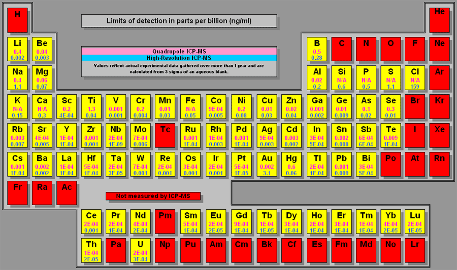
ICP-MS_elements quantifiables_EA.gif

Ablation laser couplé avec l'ICP-MS

ICP-OES_Agilent.webp

ICP-OES

Choix techniques Agilent_ICP-OES vs MS_e

Spectroscopie d'émission optique/plasma à couplage inductif
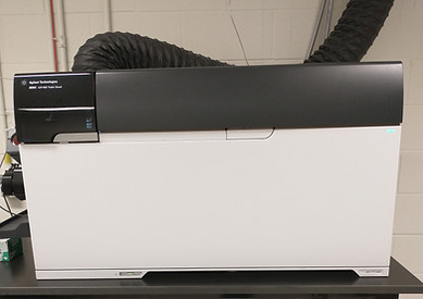
ICP-MS et pompe.jpg

Éléments chimiques détectables (en jaune)

ICP-MS_elements quantifiables_EA.gif

Spectrométrie de masse/plasma à couplage inductif (ICP-MS)

FLASH-2000-Analyzer.jpg

Pourcentages C, H, N et S

CHNS analyzer_results.jpg

Analyse CHNS (Carbone-Hydrogène-Azote-Soufre)

image.png
Survey-HR batteries_XPS Kratos.jpg

Spectroscopie photoélectronique induite par rayons X (XPS)

FTIR spectro_chimie (1).png
Spectres IR_transmission.jpg

Spectroscopie infrarouge (FTIR)

raman-1.jpg

Images Raman

Applications for Raman.jpg

Microscopie Raman confocale

Cary-Eclipse-Fluorescence-Spectrophotometer.jpg
Spectre fluo_Ex-Em_Agilent.jpg

Excitation - Émission

Spectrofluorimétrie

Cary-7000_Image.webp
NIR-UV-Visible-Spectres cuvettes_Jasco.webp

Spectroscopie NIR-UV-Visible

EPR Bruker.jpeg

Détection des électrons non appariés

epr-example-spectra_Bruker.jpeg

Résonance paramagnétique électronique (EPR ou ESR)

Contact angle Dataphysics OCA50_REGAL.jpg
Goniomètre-MT-3.jpg

Mouillabilité de la surface

Goniomètre

image.png

Taille des nanoparticules

DLS data_example Malvern.png

Diffusion dynamique de la lumière (DLS)

DSC_CERMA.jpg
Image DSC.jpg

Calorimétrie différentielle à balayage (DSC)

TGA_CERMA.jpg
TGA resultats.jpeg

Analyse thermogravimétrique (TGA)

RMN 400_chimie.jpg
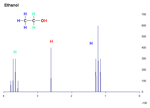
RMN spectra_ethanol 1H_Bruker.gif

Résonance magnétique nucléaire (RMN)

image.png
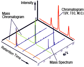
Chromato et MS_Shimadzu.gif

HPLC-MS

Chromatographie en phase liquide/spectrométrie de masse

GC-MS_chimie.jpg
Spectre MS_GC-MS.jpg

GC-MS

Chromatographie en phase gazeuse/spectrométrie de masse

Pyrolyseur.jpg
Py-GC-MS_polymeres_Frontier Lab.jpg

Pyrolyse-GC-MS

GPC_chimie.jpg
Chromatogramme_CERMA.jpg

Chromatographie d'exclusion stérique (SEC ou GPC)

Caractérisation structurale et microstructurale

DRX plus clair_gch.jpg
DRX spectra_lowT vs highT_Malvern PANalytical Empyrean.jpg

Diffraction des rayons X (DRX)

Micro-tomodensitometre_CRMR.jpg
CT scan_bone applications_Bruker.jpeg

Micro-tomodensitomètre (XRM)

Caractérisation mécanique

Nanoindenter-bruker.png

Nanoindentation

Image nanoindentation_MOD.jpg
DMA analyzers_Anton Paar.jpg
DMA thermogram_Anton Paar.jpg

Analyse thermomécanique dynamique (DMA)

Vous ne trouvez pas la technique dont vous avez besoin ci-haut? Contactez-nous et nous vous aiderons à la trouver!

bottom of page